オトガイ孔
| 骨: オトガイ孔 | |
|---|---|
 Mandible. Outer surface. Side view. (Mental foramen visible at left.) | |
| 名称 | |
| 日本語 | オトガイ孔 |
| 英語 | Mental foramen |
| ラテン語 | foramen mentale |
| 関連情報 | |
| グレイ解剖学 | 書籍中の説明(英語) |
| テンプレートを表示 | |
オトガイ孔(-こう)は下顎骨の前面にある孔。下顎管の前端で[1]、オトガイ神経とオトガイ動脈、オトガイ静脈が通る。
位置・形態
オトガイ孔は通常成長と共に後方に移動し、成人では半数以上が下顎第二小臼歯の位置にある[2]。
無歯顎や大臼歯・小臼歯欠損の人ではオトガイ孔の位置は低くなる[3][4]。
通常、オトガイ孔は左右1対であるが、3.5%から24.6%の割合で複数のオトガイ孔が認められる事があり、通常大きさは揃っておらず一つの大きなオトガイ孔とその他の副オトガイ孔となる[5]。左右とも副オトガイ孔を認めるものは少ない[5]。

関連画像
-
 Side view of the skull.
 -
 The skull from the front.
 -
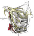 Distribution of the maxillary and mandibular nerves, and the submaxillary ganglion.
 -
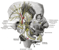 Mandibular division of the trifacial nerve.
 -
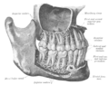 The permanent teeth, viewed from the right.


脚注
[脚注の使い方]
- ^ 原著:森於菟、改訂:森富「骨学II.頭蓋 B.顔面骨頭蓋骨 4.下顎骨」『分担解剖学1』(第11版第20刷)金原出版、東京都文京区、2000年11月20日、78-94頁。ISBN 978-4-307-00341-4。
- ^ 竹之下康治「オトガイ孔の加齢的変化 位置および開放方向の変化について」(PDF)『日本口腔外科学会雑誌』第24巻第3号、日本口腔外科学会、1978年6月、481-487頁、doi:10.5794/jjoms.24.481、ISSN 0021-5163、NAID 40003965097、JOI:JST.Journalarchive/jjoms1967/24.481、2011年11月10日閲覧。
- ^ Soikkonen K, Wolf J, Ainamo A, Xie Q. (November 1995). “Changes in the position of the mental foramen as a result of alveolar atrophy”. J Oral Rehabil. 22 (11): 831–3. doi:10.1111/j.1365-2842.1995.tb00230.x. PMID 8558356.
- ^ 関口洋介「有歯顎および大・小臼歯欠損顎におけるオトガイ孔の差異について」(PDF)『日本口腔科学会雑誌』第22巻第3号、日本口腔科学会、1973年1月、351-355頁、ISSN 0029-0297、JOI:JST.Journalarchive/stomatology1952/22.351、2011年11月10日閲覧。
- ^ a b 澤裕一郎、熊澤友子、滝本明、馬杉亮彦、川野大、野村明日香「3D-CT画像による副オトガイ孔の発現頻度に関する検討」(PDF)『日本口腔外科学会雑誌』第50巻第6号、日本口腔外科学会、2004年6月、408-411頁、doi:10.5794/jjoms.50.408、ISSN 0021-5163、NAID 10018619915、JOI:JST.Journalarchive/jjoms1967/50.408、2011年11月10日閲覧。
外部リンク
- cranialnerves at The Anatomy Lesson by Wesley Norman (Georgetown University) (V)
- Anatomy diagram: 34256.000-1 at Roche Lexicon - illustrated navigator, Elsevier
- Anatomy diagram: 34256.000-2 at Roche Lexicon - illustrated navigator, Elsevier
- SUNY - lateral view
- SUNY - frontal view
- Diagram at uni-mainz.de
| 典拠管理データベース |
|
|---|
- 表示
- 編集


