プラスミノーゲンアクチベーターインヒビター2
| SERPINB2 | |||||||||||||||||||||||||
|---|---|---|---|---|---|---|---|---|---|---|---|---|---|---|---|---|---|---|---|---|---|---|---|---|---|
 | |||||||||||||||||||||||||
| 識別子 | |||||||||||||||||||||||||
| 記号 | SERPINB2, HsT1201, PAI, PAI-2, PAI2, PLANH2, serpin family B member 2 | ||||||||||||||||||||||||
| 外部ID | OMIM: 173390 MGI: 97609 HomoloGene: 20571 GeneCards: SERPINB2 | ||||||||||||||||||||||||
| |||||||||||||||||||||||||
| |||||||||||||||||||||||||
| |||||||||||||||||||||||||
| |||||||||||||||||||||||||
| オルソログ | |||||||||||||||||||||||||
| 種 | ヒト | マウス | |||||||||||||||||||||||
| Entrez |
|
| |||||||||||||||||||||||
| Ensembl |
|
| |||||||||||||||||||||||
| UniProt |
|
| |||||||||||||||||||||||
| RefSeq (mRNA) |
|
| |||||||||||||||||||||||
| RefSeq (タンパク質) |
|
| |||||||||||||||||||||||
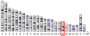
| 場所 (UCSC) | Chr 18: 63.87 – 63.9 Mb | Chr 18: 107.44 – 107.46 Mb | |||||||||||||||||||||||
| PubMed検索 | [3] | [4] | |||||||||||||||||||||||
| ウィキデータ | |||||||||||||||||||||||||
| |||||||||||||||||||||||||


プラスミノーゲンアクチベーターインヒビター2(英: plasminogen activator inhibitor 2、PAI-2)、SERPINB2または胎盤性PAI(placental PAI)は、セルピンスーパーファミリーに属するセリンプロテアーゼインヒビターであり、t-PAやウロキナーゼを不活化する血液凝固因子である。PAI-2は大部分の細胞、特に単球/マクロファージに存在している。PAI-2はグリコシル化された約60 kDaの細胞外型、そして約43 kDaの細胞内型の2つの形態で存在する。

PAI-2は胎盤から分泌されるため、妊娠中にのみ検出可能なレベルで血中に存在し、また妊娠中の血栓傾向の増大に部分的に寄与している可能性がある。PAI-2のタンパク質内部に存在するシグナルペプチドは効率が低いため、発現したPAI-2の大部分は分泌されず細胞内にとどまる。
相互作用
PAI-2はさまざまな細胞内・細胞外タンパク質に結合することが報告されている。PAI-2の生理的機能がウロキナーゼなどの細胞外プロテアーゼの阻害であるのかどうか、そしてPAI-2に細胞内での活性が存在するかどうかに関しては議論がある。PAI-2の生理的機能の少なくとも1つは、獲得免疫の調節と関係したものである可能性がある[5]。
構造と多量体化
他のセルピンと同様、PAI-2には3つのβシート(A、B、C)と9本のαヘリックス(hA–hI)が存在する[6][7]。hCとhDを連結する33アミノ酸を欠失したPAI-2変異体の構造が解かれている。このCDループは特に柔軟性が高く安定化が困難であり、分子内でのジスルフィド結合時に最大で54 Åも移動することが知られている[8]。他の特筆すべきモチーフとしては、379–383番に位置するRCL(reactive center loop)、N末端の疎水性シグナル配列などがある。

PAI-1とPAI-2は阻害標的が類似しているにもかかわらず、系統学的には遠い関係にある。PAI-2はオボアルブミン関連セルピンファミリーのメンバーであり、ニワトリのオボアルブミンと遺伝学的に類似している。PAI-2は哺乳類におけるオボアルブミンに近縁のホモログである[9]。オボアルブミンとPAI-2はどちらも非切断型の分泌シグナルペプチドによって分泌されるが、PAI-2の分泌はオボアルブミンと比較してかなり非効率である[10]。
PAI-2は単量体型、多量体型(不活性状態)、そしてpolymerigenic formと呼ばれる中間体の3つの状態で存在する。多量化はいわゆる"loop-sheet"機構によって行われ、ある分子のRCLが隣接する分子のβシートAへ逐次挿入される。この過程はPAI-2がpolymerigenic formの状態のときに選択的に生じる。Polymerigenic formはCys79(CDループに位置する)とCys161の間のジスルフィド結合によって安定化されている[11]。単量体型のPAI-2ではCDループはかなり離れた位置にあり、Cys161と十分に近接してジスルフィド結合を形成するためには54 Åの距離を移動する必要がある。しかしながらCDループは極めて柔軟性が高いため、単量体型とpolymerigenic formは完全に相互変換可能であり、タンパク質の酸化還元環境の変化によってどちらの状態が好まれるかが変化する[8]。PAI-2の多量体化は、胎盤細胞の細胞質基質など、生理的条件下で自発的に生じる[12]。細胞質基質ではPAI-2は単量体型である傾向にあるが、分泌と関連したオルガネラ(細胞質基質よりも酸化的傾向がある)ではPAI-2の多量体化はより生じやすい[11]。こうした理由により、PAI-2は環境の酸化還元電位を検知して応答している可能性があると考えられている[8]。
機構
PAI-2は自殺阻害(英語版)機構を利用する。この機構はセルピンでは一般的であり、t-PAやウロキナーゼを不可逆的に不活性化する[6]。まず、標的となるセリンプロテアーゼがPAI-2にドッキングし、RCLのArg380とThr381の間の切断を触媒する。この時点では、2つの結果が生じる可能性がある。プロテアーゼが解離し、不活性なPAI-2が生じる。または、プロテアーゼはPAI-2と永続的な共有結合複合体を形成し、酵素活性を失う。この複合体ではプロテアーゼは大きく変形している。
生物学的機能
細胞外の(グリコシル化された)PAI-2は線維素溶解(英語版)を調節する機能を果たすが、この阻害がPAI-2の主要な機能であるのかどうかは明確ではない。PAI-2は主に細胞内に存在し、その分泌シグナルペプチドは比較的効率が低い。このシグナル配列の変異によって分泌効率は大きく高まることから、この効率の低さはおそらく進化的デザインによるものである[10]。PAI-2は通常は成人の血漿には検出されず、妊娠中、骨髄単球性白血病といったケースや、歯肉溝滲出液中にのみ検出される。さらに、PAI-2による阻害はPAI-1と比較してオーダーが異なるほど遅い(二次反応速度定数に基づく)[13]。一方で、PAI-2の細胞内での詳細な役割に関する結論が得られているわけでもない。
PAI-2は妊娠中や免疫応答時にアップレギュレーションされる。妊娠時にはPAI-2は特に脱落膜(英語版)や羊水中に存在し、そこで膜を分解から保護し、胎児や子宮の組織のリモデリングを補助している[14]。PAI-2は線維素溶解の調節過程においてPAI-1を補助し、また血栓症のリスクを高めるPAI-1の過剰発現を防いでいる可能性がある[14][15]。妊娠期間に血漿中のPAI-2濃度はほぼ検出不可能なレベルから250 ng/mLにまで上昇する(その大部分がグリコシル化型でである)[13]。
免疫細胞の中では、PAI-2を産生しているのは主にマクロファージであり、B細胞やT細胞が多くを産生することはない[16]。PAI-2は炎症応答や感染に関与しており、IgG2やII型インターフェロンを分泌するT細胞のダウンレギュレーションに関与している可能性がある[16]。
PAI-2をコードするSERPINB2遺伝子は18番染色体(英語版)にBCL2や他のセルピン遺伝子と近接して位置しているため、アポトーシスにおける役割の研究が行われている。しかしながら、決定的なエビデンスは今のところ得られていない[13][17]。近年の研究では、PAI-2がp53の直接下流の標的で活性化因子であり、p21を直接的に安定化している可能性が示唆されている。さらに、老化線維芽細胞ではPAI-2の発現が上昇していることから、若い線維芽細胞の成長停止に関与している可能性がある[18]。
がんにおける役割
PAI-2は腫瘍促進効果と抑制効果の双方を有する可能性があるため、がんの成長や転移における役割は複雑である。特筆すべきこととして、関連する組織ではなく腫瘍細胞自体によるPAI-2の高発現ががん細胞の成長に影響を及ぼす[19]。がん細胞はマイクロパーティクルと呼ばれる小胞を介してPAI-2の放出を促進している可能性がある[19]。
PAI-2は、腫瘍に対して致死的影響を発揮する場合のある、プラスミン誘発性の細胞死からがん細胞を保護する。この保護効果は転移性脳腫瘍で特に顕著である。転移性脳腫瘍ではPAI-2やニューロセルピン(英語版)が高レベルで発現している傾向があり、その成長はPAI-2のノックアウトによって部分的に阻害される[20]。PAI-2は腫瘍細胞で高発現しているため、メラノーマ細胞の血行性転移による拡散の追跡や研究にも利用されている[21]。
PAI-2の発現は脳への転移を促進する場合があるが、他のケースではPAI-2の高発現が肺やその他の器官への転移を有意に低下させる[19][22]。そのため、転移におけるPAI-2の影響はがんの種類や体内の部位に依存している可能性がある。
出典
- ^ a b c GRCh38: Ensembl release 89: ENSG00000197632 - Ensembl, May 2017
- ^ a b c GRCm38: Ensembl release 89: ENSMUSG00000062345 - Ensembl, May 2017
- ^ Human PubMed Reference:
- ^ Mouse PubMed Reference:
- ^ “The role of SerpinB2 in immunity”. Critical Reviews in Immunology 31 (1): 15–30. (2011). doi:10.1615/critrevimmunol.v31.i1.20. PMID 21395508.
- ^ a b “An overview of the serpin superfamily”. Genome Biology 7 (5): 216. (2006). doi:10.1186/gb-2006-7-5-216. PMC 1779521. PMID 16737556. https://www.ncbi.nlm.nih.gov/pmc/articles/PMC1779521/.
- ^ “Plasminogen activator inhibitor-2 is highly tolerant to P8 residue substitution--implications for serpin mechanistic model and prediction of nsSNP activities”. Journal of Molecular Biology 353 (5): 1069–80. (November 2005). doi:10.1016/j.jmb.2005.09.008. PMID 16214170.
- ^ a b c “Structural bases of the redox-dependent conformational switch in the serpin PAI-2”. Journal of Molecular Biology 344 (5): 1359–68. (December 2004). doi:10.1016/j.jmb.2004.10.010. PMID 15561148.
- ^ “Structure of the gene for human plasminogen activator inhibitor-2. The nearest mammalian homologue of chicken ovalbumin”. The Journal of Biological Chemistry 264 (10): 5495–502. (April 1989). doi:10.1016/S0021-9258(18)83572-4. PMID 2494165.
- ^ a b “Functional activity of eukaryotic signal sequences in Escherichia coli: the ovalbumin family of serine protease inhibitors”. Journal of Molecular Biology 335 (2): 437–53. (January 2004). doi:10.1016/j.jmb.2003.10.076. PMID 14672654.
- ^ a b “A redox-sensitive loop regulates plasminogen activator inhibitor type 2 (PAI-2) polymerization”. The EMBO Journal 22 (8): 1753–61. (April 2003). doi:10.1093/emboj/cdg178. PMC 154470. PMID 12682008. https://www.ncbi.nlm.nih.gov/pmc/articles/PMC154470/.
- ^ “Intracellular polymerization of the serpin plasminogen activator inhibitor type 2”. The Journal of Biological Chemistry 271 (17): 10048–53. (April 1996). doi:10.1074/jbc.271.17.10048. PMID 8626560.
- ^ a b c “Biological and clinical aspects of plasminogen activator inhibitor type 2”. Blood 86 (11): 4007–24. (December 1995). doi:10.1182/blood.v86.11.4007.bloodjournal86114007. PMID 7492756.
- ^ a b “Significance of the plasminogen activator inhibitor of placental type (PAI-2) in pregnancy”. Seminars in Thrombosis and Hemostasis 24 (5): 431–5. (1998). doi:10.1055/s-2007-996035. PMID 9834009.
- ^ “Rebound elevation of fibronectin after tissue injury and ischemia: role of fibronectin synthesis”. The American Journal of Physiology 263 (4 Pt 1): G437–45. (October 1992). doi:10.1152/ajpgi.1992.263.4.G437. PMID 1415704.
- ^ a b “A physiological function of inflammation-associated SerpinB2 is regulation of adaptive immunity”. Journal of Immunology 184 (5): 2663–70. (March 2010). doi:10.4049/jimmunol.0902187. PMID 20130210.
- ^ “Forty years later and the role of plasminogen activator inhibitor type 2/SERPINB2 is still an enigma”. Seminars in Thrombosis and Hemostasis 37 (4): 395–407. (June 2011). doi:10.1055/s-0031-1276589. PMID 21805446.
- ^ “The serine protease inhibitor serpinB2 binds and stabilizes p21 in senescent cells”. Journal of Cell Science 130 (19): 3272–3281. (October 2017). doi:10.1242/jcs.204974. PMID 28794016.
- ^ a b c “Tumor cell-expressed SerpinB2 is present on microparticles and inhibits metastasis”. Cancer Medicine 3 (3): 500–13. (June 2014). doi:10.1002/cam4.229. PMC 4101741. PMID 24644264. https://www.ncbi.nlm.nih.gov/pmc/articles/PMC4101741/.
- ^ “Serpins promote cancer cell survival and vascular co-option in brain metastasis”. Cell 156 (5): 1002–16. (February 2014). doi:10.1016/j.cell.2014.01.040. PMC 3988473. PMID 24581498. https://www.ncbi.nlm.nih.gov/pmc/articles/PMC3988473/.
- ^ “Imaging of Angiotropism/Vascular Co-Option in a Murine Model of Brain Melanoma: Implications for Melanoma Progression along Extravascular Pathways”. Scientific Reports 6: 23834. (April 2016). Bibcode: 2016NatSR...623834B. doi:10.1038/srep23834. PMC 4822155. PMID 27048955. https://www.ncbi.nlm.nih.gov/pmc/articles/PMC4822155/.
- ^ “Overexpression of plasminogen activator inhibitor 2 in human melanoma cells inhibits spontaneous metastasis in scid/scid mice”. Proceedings of the National Academy of Sciences of the United States of America 92 (1): 205–9. (January 1995). Bibcode: 1995PNAS...92..205M. doi:10.1073/pnas.92.1.205. PMC 42846. PMID 7816818. https://www.ncbi.nlm.nih.gov/pmc/articles/PMC42846/.
関連文献
- “Microsequences of 145 proteins recorded in the two-dimensional gel protein database of normal human epidermal keratinocytes”. Electrophoresis 13 (12): 960–9. (December 1992). doi:10.1002/elps.11501301199. PMID 1286667.
- “Inhibition of receptor-bound urokinase by plasminogen-activator inhibitors”. The Journal of Biological Chemistry 265 (17): 9904–8. (June 1990). doi:10.1016/S0021-9258(19)38757-5. PMID 2161846.
- “The receptor for urokinase type plasminogen activator polarizes expression of the protease to the leading edge of migrating monocytes and promotes degradation of enzyme inhibitor complexes”. The Journal of Cell Biology 111 (2): 783–92. (August 1990). doi:10.1083/jcb.111.2.783. PMC 2116194. PMID 2166055. https://www.ncbi.nlm.nih.gov/pmc/articles/PMC2116194/.
- “Chromosomal organization and localization of the human urokinase inhibitor gene: perfect structural conservation with ovalbumin”. Genomics 6 (1): 159–67. (January 1990). doi:10.1016/0888-7543(90)90461-3. PMID 2303256.
- “Endotoxin-induced production of plasminogen activator inhibitor by human monocytes is autonomous and can be inhibited by lipid X”. Blood 73 (8): 2188–95. (June 1989). doi:10.1182/blood.V73.8.2188.2188. PMID 2471561.
- “Structure of the gene for human plasminogen activator inhibitor-2. The nearest mammalian homologue of chicken ovalbumin”. The Journal of Biological Chemistry 264 (10): 5495–502. (April 1989). doi:10.1016/S0021-9258(18)83572-4. PMID 2494165.
- “Isolation of multiple types of plasminogen activator inhibitors from vascular smooth muscle cells”. Thrombosis and Haemostasis 61 (3): 517–21. (June 1989). doi:10.1055/s-0038-1646626. PMID 2799763.
- “Plasminogen activator inhibitor 2. Isolation and characterization of the promoter region of the gene”. Biochemical and Biophysical Research Communications 156 (1): 383–8. (October 1988). doi:10.1016/S0006-291X(88)80852-0. PMID 2845977.
- “cDNA cloning and expression in Escherichia coli of a plasminogen activator inhibitor from human placenta”. The Journal of Biological Chemistry 262 (8): 3718–25. (March 1987). doi:10.1016/S0021-9258(18)61414-0. PMID 3029122.
- “Cloning and expression of a cDNA coding for a human monocyte-derived plasminogen activator inhibitor”. Proceedings of the National Academy of Sciences of the United States of America 85 (4): 985–9. (February 1988). Bibcode: 1988PNAS...85..985A. doi:10.1073/pnas.85.4.985. PMC 279685. PMID 3257578. https://www.ncbi.nlm.nih.gov/pmc/articles/PMC279685/.
- “Plasminogen activator inhibitor 2: regulation of gene transcription during phorbol ester-mediated differentiation of U-937 human histiocytic lymphoma cells”. Molecular and Cellular Biology 7 (12): 4564–7. (December 1987). doi:10.1128/mcb.7.12.4564. PMC 368144. PMID 3325828. https://www.ncbi.nlm.nih.gov/pmc/articles/PMC368144/.
- “Human monocyte Arg-Serpin cDNA. Sequence, chromosomal assignment, and homology to plasminogen activator-inhibitor”. The Journal of Experimental Medicine 166 (1): 77–94. (July 1987). doi:10.1084/jem.166.1.77. PMC 2188630. PMID 3496414. https://www.ncbi.nlm.nih.gov/pmc/articles/PMC2188630/.
- “Plasminogen activator inhibitor type 2 inhibits tumor necrosis factor alpha-induced apoptosis. Evidence for an alternate biological function”. The Journal of Biological Chemistry 270 (46): 27894–904. (November 1995). doi:10.1074/jbc.270.46.27894. PMID 7499264.
- “Plasminogen-activator inhibitor type 2 (PAI-2) is a spontaneously polymerising SERPIN. Biochemical characterisation of the recombinant intracellular and extracellular forms”. European Journal of Biochemistry 218 (3): 1071–82. (December 1993). doi:10.1111/j.1432-1033.1993.tb18467.x. PMID 7506655.
- “Plasminogen activator inhibitor type 2: an intracellular keratinocyte differentiation product that is incorporated into the cornified envelope”. Experimental Cell Research 217 (1): 65–71. (March 1995). doi:10.1006/excr.1995.1064. PMID 7867722.
- “Microglia express the type 2 plasminogen activator inhibitor in the brain of control subjects and patients with Alzheimer's disease”. Neuroscience Letters 164 (1–2): 233–5. (December 1993). doi:10.1016/0304-3940(93)90899-V. PMID 8152607.
- “Processing of complex between urokinase and its type-2 inhibitor on the cell surface. A possible regulatory mechanism of urokinase activity”. FEBS Letters 323 (3): 279–84. (June 1993). doi:10.1016/0014-5793(93)81357-6. PMID 8388810.
- “Cytoplasmic antiproteinase 2 (PI8) and bomapin (PI10) map to the serpin cluster at 18q21.3”. Genomics 43 (3): 321–8. (August 1997). doi:10.1006/geno.1997.4827. PMID 9268635.
- “DNase I hypersensitive sites in the 5' flanking region of the human plasminogen activator inhibitor type 2 (PAI-2) gene are associated with basal and tumor necrosis factor-alpha-induced transcription in monocytes”. European Journal of Biochemistry 256 (3): 550–9. (September 1998). doi:10.1046/j.1432-1327.1998.2560550.x. PMID 9780231.
- “Expression and localization of the urokinase-type plasminogen activator receptor (uPAR) in the human placenta”. The Kobe Journal of Medical Sciences 44 (1): 31–43. (February 1998). PMID 9846056.
関連項目
外部リンク
- ペプチダーゼとそのインヒビターに関するMEROPSオンラインデータベース: I04.007
- Plasminogen Activator Inhibitor 2 - MeSH・アメリカ国立医学図書館・生命科学用語シソーラス(英語)


